Brain Imaging.
Visualized. Analyzed.
NIfTI brain viewer with atlas overlays, FSL integration, MELD analysis, and PACS import. Explore 3D volumes right in your browser.
Get Started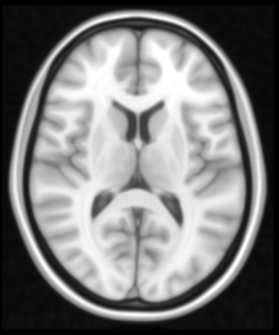 Axial
Axial
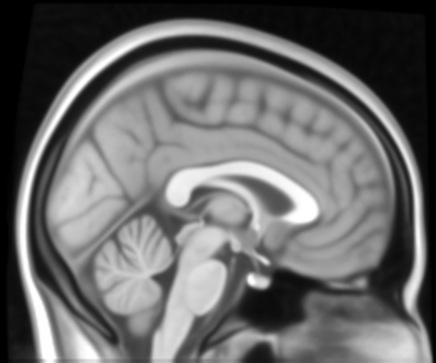 Sagittal
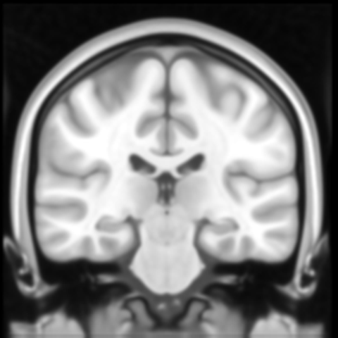 Coronal
Built for Neuroscience
Specialized tools for brain imaging — from clinical neurology to research pipelines.
NIfTI Viewer
Load NII / NII.GZ volumes, navigate axial, sagittal, coronal slices. Adjust contrast, window, colormap in real time.
Atlas Overlays
MNI152 template, Harvard-Oxford cortical/subcortical atlases. Toggle regions, adjust opacity, identify structures.
FSL Integration
Run FSL pipelines server-side. BET extraction, FLIRT registration, FAST segmentation — results rendered instantly.
MELD Analysis
Automated focal cortical dysplasia detection on brain MRI. Calibrated on 1.5T Siemens. T1, T1+FLAIR supported.
PACS Import
Import DICOM series from your PACS, auto-convert to NIfTI. Study worklist with patient metadata and volume management.
Multi-volume
Load multiple volumes per study. Overlay masks, compare time points, toggle between sequences — all in one canvas.
Three simple steps
Upload NIfTI
Drag & drop NII.GZ files or import from PACS. Auto-convert DICOM to NIfTI.
Visualize & Overlay
Navigate slices, apply atlases, run AI analysis. See results in real time.
Analyze & Export
Run MELD, FSL pipelines. Export results, generate reports.
Ready to explore?
Sign in to access NeuroView, or create a free account.
 Continue with Google