Bone age assessment
AI-powered bone age estimation from hand radiographs, with chronological comparison and optional PDF report
Research tool — not a regulated medical device. Their results are not a diagnosis and can be wrong. Every result must be reviewed by a qualified clinician, who remains responsible for the clinical decision.
What it does
Estimates skeletal maturity (bone age) from a paediatric hand-and-wrist X-ray using a deep neural network, and reports the predicted age against chronological age.
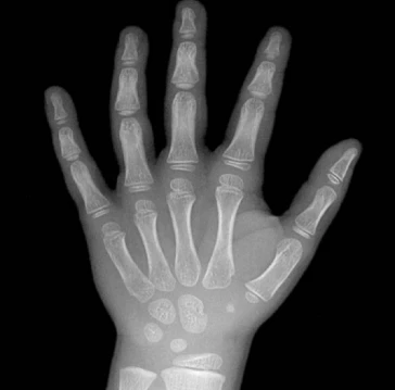
AI result
Predicted Bone Age
4 years 11 months
Chronological Age
5 years
Difference
−1 month
Within normal range
What is bone age and why does it matter?
Bone age reflects the biological maturity of the skeleton, assessed from ossification centres visible on a hand X-ray. Comparing bone age with chronological age helps clinicians evaluate whether a child's skeletal development is appropriate, advanced, or delayed relative to their peers.
Bone age assessment is used in paediatric endocrinology to investigate growth disorders, precocious or delayed puberty, and to monitor treatment with growth hormone. It is also used to estimate adult height potential and to evaluate children with short or tall stature.
Greulich–Pyle and TW3 atlases
The most widely used reference for manual bone age assessment is the Greulich–Pyle (GP) atlas, first published in 1950 and revised in 1959. It contains standard radiographs of the left hand and wrist for boys and girls at various ages, allowing a radiologist to compare the patient's film with the closest matching standard and assign a skeletal age.
The Tanner–Whitehouse 3 (TW3) method provides a more systematic alternative: each of 13 bones of the hand and wrist is scored individually, and the scores are summed to derive a bone age. TW3 is considered more reproducible than the GP atlas and is preferred in research settings. This tool uses an AI model trained on hand radiographs and validated against these established references.